
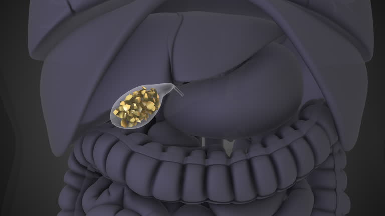
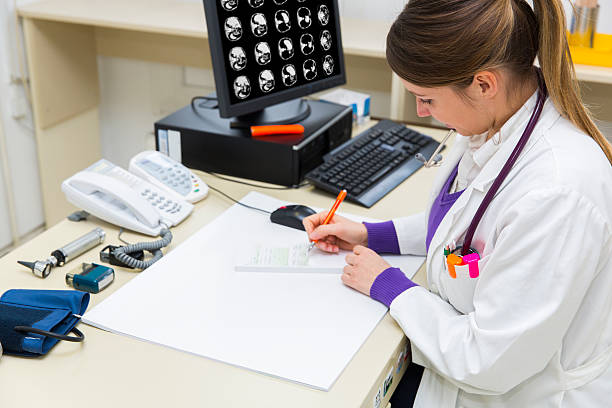
Cholecystitis, the inflammation of the gallbladder, can lead to severe pain and discomfort. Diagnosing Cholecystitis often involves a combination of tools and techniques, and the stethoscope plays an essential role in the initial evaluation. It helps doctors identify abnormal sounds in the abdomen, which can indicate underlying issues related to the gallbladder.
In this article, we will dive deeper into how stethoscopes are used alongside other diagnostic methods, including laboratory tests, imaging, and clinical guidelines, to confirm and treat cholecystitis.
How Stethoscopes Help in Diagnosing Cholecystitis?
When a patient presents with symptoms like abdominal pain, fever, or nausea, a doctor will begin with a physical examination. Using a stethoscope, the doctor listens for abnormal bowel sounds in the abdomen.
Key Observations Made with a Stethoscope:
- Reduced Bowel Sounds: A lack of gurgling noises may indicate bowel paralysis or other complications.
- Localized Sounds: Abnormal noises in the upper right abdomen, where the gallbladder is located, may suggest inflammation.
- Associated Symptoms: The stethoscope examination helps guide further tests, especially if pain increases during palpation.
While the stethoscope alone cannot confirm cholecystitis, it is an important first step in identifying the problem.
Laboratory Tests for Cholecystitis
Laboratory tests for cholecystitis are vital in confirming the diagnosis after an initial examination. Blood tests reveal the presence of infection or inflammation and help pinpoint complications.
Common Blood Tests:
- White Blood Cell Count (WBC): Elevated levels indicate infection or inflammation.
- Liver Function Tests (LFTs): High levels of enzymes like ALT, AST, or bilirubin suggest bile duct blockage.
- C-reactive Protein (CRP): An increase signals inflammation, which is common in acute cases.
These tests provide insights into the severity of the condition and guide treatment decisions.
Imaging Techniques and Gold Standard Tests
Imaging studies confirm the presence of gallstones or inflammation. The gold standard test for cholelithiasis (gallstones) is an ultrasound, which is highly effective in detecting stones or bile duct blockages.
Comparison of Imaging Methods:
| Imaging Test | Purpose | Advantages |
| Ultrasound | Detects gallstones and inflammation. | Non-invasive, reliable, and widely used. |
| CT Scan | Visualizes complications or perforations. | Provides detailed images of the abdomen. |
| HIDA Scan | Assesses gallbladder function. | Diagnoses bile flow problems. |
For acute cholecystitis, ultrasound remains the preferred method due to its accuracy and availability.
Tokyo Guidelines for Diagnosing Cholecystitis
The Tokyo Guidelines for Cholecystitis outline a systematic approach for diagnosing and managing this condition. These guidelines combine three key criteria:
- Clinical Symptoms: Fever, upper right abdominal pain, and nausea.
- Laboratory Findings: Elevated WBC, liver enzymes, and CRP levels.
- Imaging Results: Confirmation of gallstones, thickened gallbladder walls, or fluid around the gallbladder.
Following these guidelines ensures accurate diagnosis and timely treatment, reducing complications.
Gold Standard Treatment for Cholecystitis
Once diagnosed, the gold standard treatment for cholecystitis is cholecystectomy, the surgical removal of the gallbladder. This procedure can be performed using two methods:
- Laparoscopic Cholecystectomy: A minimally invasive surgery with faster recovery times.
- Open Cholecystectomy: Used in complex cases with complications like perforation or severe infection.
Early surgery prevents the condition from becoming chronic or leading to severe complications.
Differential Diagnosis of Cholecystitis
Symptoms of cholecystitis can mimic other conditions. To ensure proper treatment, doctors consider the differential diagnosis of cholecystitis, which includes:
- Pancreatitis: Inflammation of the pancreas.
- Peptic Ulcer Disease: Sores in the stomach lining.
- Irritable Bowel Syndrome (IBS): Chronic abdominal discomfort.
Imaging and laboratory tests help rule out these conditions.
Cholecystitis Bloods: What They Reveal
Blood tests, often referred to as cholecystitis blood, offer critical information about the condition.
Key Indicators:
- High White Blood Cells: Signal infection.
- Elevated Bilirubin Levels: Indicates bile duct blockage.
- Abnormal Liver Enzymes: Suggest gallbladder or bile duct dysfunction.
These findings guide doctors toward the appropriate treatment plan.
Chronic Cholecystitis: A Long-Term Issue
Unlike the sudden onset of acute cholecystitis, chronic cholecystitis develops over time due to recurring inflammation.
Symptoms:
- Persistent abdominal pain, especially after eating fatty foods.
- Nausea and bloating.
- Increased risk of gallbladder dysfunction or cancer.
Treatment often involves removing the gallbladder to prevent further complications.
Conclusion: The Role of Stethoscopes
The role of stethoscopes in diagnosing cholecystitis is foundational. They provide initial clues that guide further testing, like imaging and blood work. Combined with tools like the gold standard test for cholelithiasis (ultrasound) and treatment options such as cholecystectomy, early diagnosis ensures better outcomes.
By understanding how stethoscopes and other methods work together, you can appreciate the value of these tools in managing conditions like cholecystitis effectively.