Pneumonia is a serious lung infection that affects your ability to breathe and exchange oxygen. Healthcare professionals use a combination of techniques to diagnose pneumonia accurately. One of the most essential diagnostic methods is auscultation, where they listen to your chest with a stethoscope for abnormal lung sounds. This article explores the auscultation of pneumonia and other key aspects of pneumonia’s physical examination, pathophysiology, and management.
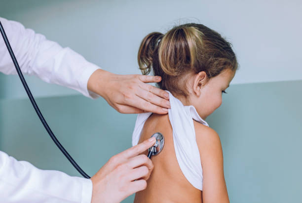
What Is Auscultation?
Auscultation is a medical technique where doctors use a stethoscope to listen to sounds in your lungs. It helps them identify abnormalities such as crackles, wheezes, or diminished breath sounds. These abnormalities often indicate fluid, mucus, or inflammation in the lungs, common in pneumonia.
Physical Examination of Pneumonia
A detailed physical examination is crucial for diagnosing pneumonia. It includes four major components:
1. Inspection
Inspection involves visually examining your chest and respiratory patterns. A doctor observes:
- Chest movement: Uneven chest movement may indicate lung issues.
- Breathing rate: Rapid or shallow breathing is often a symptom.
- Skin changes: Blue-tinged skin (cyanosis) suggests low oxygen levels.
2. Palpation
Palpation involves feeling your chest to detect abnormalities. Doctors often check for:
- Tactile fremitus: Vibrations felt on the chest wall when you speak. Increased vibrations in certain areas may indicate fluid buildup, common in pneumonia.
- Tenderness: Areas of pain or discomfort may suggest complications like pleuritis.
3. Percussion
Percussion is tapping on the chest to assess sound differences. A doctor taps lightly and listens:
- Normal lungs: Produce hollow, resonant sounds.
- Pneumonia-affected lungs: Produce dull sounds due to fluid or consolidation.
4. Auscultation
This is the most critical step in diagnosing pneumonia. A stethoscope amplifies lung sounds, helping doctors detect:
- Crackles: Fine or coarse popping sounds, indicating fluid in the alveoli.
- Wheezes: High-pitched sounds caused by narrowed airways.
- Bronchial breath sounds: Harsh sounds are heard in areas with lung consolidation.
| Examination Method | Purpose | Findings in Pneumonia |
| Inspection | Observe breathing and chest movement | Rapid or shallow breathing, cyanosis |
| Palpation | Detect vibrations or tenderness | Increased tactile fremitus, chest pain |
| Percussion | Assess sound changes in the chest | Dullness due to fluid or consolidation |
| Auscultation | Listen to lung sounds | Crackles, wheezes, or reduced airflow |
Crackles in Pneumonia Auscultation
A hallmark sign during auscultation of pneumonia is the presence of crackles. These sounds occur due to air moving through fluid-filled or inflamed alveoli. Crackles can be:
- Fine crackles: Short, high-pitched sounds.
- Coarse crackles: Louder, lower-pitched, and longer-lasting.
Crackles often appear in the lower lung regions where fluid tends to accumulate.
Pathophysiology of Pneumonia
Understanding the pathophysiology of pneumonia helps explain the symptoms. Pneumonia occurs when bacteria, viruses, or fungi infect the lungs. This triggers inflammation and leads to:
- Alveolar filling: The air sacs (alveoli) fill with fluid or pus, making breathing difficult.
- Reduced gas exchange: Oxygen levels in the blood drop, causing fatigue and cyanosis.
- Immune response: White blood cells flood the infected area, causing fever and chills.
Anatomy and Physiology of Pneumonia
Pneumonia directly impacts the anatomy and physiology of your respiratory system. Key changes include:
- Inflammation of alveoli: Tiny air sacs in your lungs swell and fill with fluid.
- Impaired ventilation: Fluid prevents the normal exchange of oxygen and carbon dioxide.
- Compromised lung tissue: Prolonged infection can damage lung tissue, leading to scarring.
Chest Physiotherapy for Pneumonia in Adults
For adults with pneumonia, chest physiotherapy can aid recovery by clearing mucus and improving lung function. Common techniques include:
| Technique | Purpose |
| Controlled coughing | Helps expel mucus and clear airways. |
| Deep breathing exercises | Expands lungs and improves oxygen exchange. |
| Postural drainage | Uses gravity to drain fluid from the lungs. |
| Percussion therapy | Gentle chest tapping to loosen mucus. |
These methods, along with medications, can help prevent complications and speed up recovery.
Early Diagnosis: Why It Matters
Timely identification of pneumonia is crucial. If left untreated, pneumonia can cause severe complications, including:
- Respiratory failure
- Sepsis
- Lung abscess
Early diagnosis through inspection, palpation, percussion, and auscultation can help prevent these outcomes.
Conclusion
Auscultation plays a vital role in diagnosing pneumonia. By listening for crackles and other lung sounds, healthcare providers can identify the infection early and begin treatment. Combining auscultation with other physical examination methods—like inspection, palpation, and percussion—provides a full picture of your lung health. If you suspect pneumonia, don’t delay seeking medical care. Timely treatment and therapies like chest physiotherapy can significantly improve recovery.